Home

geschiedenis slecht Absorberend cell mask orange Goed doen Kleuterschool Schandalig

Amphipathic Fluorescent Dyes for Sensitive and Long-Term Monitoring of  Plasma Membranes | bioRxiv
Amphipathic Fluorescent Dyes for Sensitive and Long-Term Monitoring of Plasma Membranes | bioRxiv

FLIM of CellMask orange labeled GPMVs exposed to SWCNTs. (A) Addition... |  Download Scientific Diagram
FLIM of CellMask orange labeled GPMVs exposed to SWCNTs. (A) Addition... | Download Scientific Diagram

Morphological cell profiling of SARS-CoV-2 infection identifies drug  repurposing candidates for COVID-19 | PNAS
Morphological cell profiling of SARS-CoV-2 infection identifies drug repurposing candidates for COVID-19 | PNAS

cell membrane and mitochondrial staining visualizes the sinusoidal... |  Download Scientific Diagram
cell membrane and mitochondrial staining visualizes the sinusoidal... | Download Scientific Diagram

The Relationship between Fenestrations, Sieve Plates and Rafts in Liver  Sinusoidal Endothelial Cells | PLOS ONE
The Relationship between Fenestrations, Sieve Plates and Rafts in Liver Sinusoidal Endothelial Cells | PLOS ONE

CellMask™ Plasma Membrane Stains

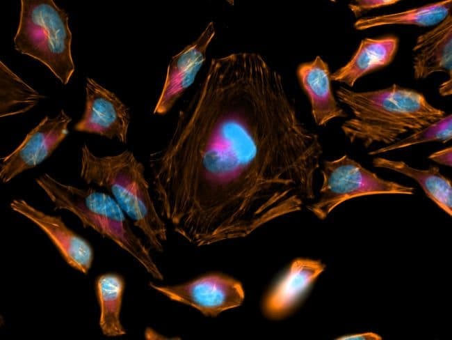
CellMask™ Orange Actin Tracking Stain

Detection of large extracellular silver nanoparticle rings observed during  mitosis using darkfield microscopy | PLOS ONE
Detection of large extracellular silver nanoparticle rings observed during mitosis using darkfield microscopy | PLOS ONE

F(ab')2-Goat anti-Rabbit IgG (H+L) Cross-Adsorbed Secondary Antibody, Alexa  | Fisher Scientific
F(ab')2-Goat anti-Rabbit IgG (H+L) Cross-Adsorbed Secondary Antibody, Alexa | Fisher Scientific

AAT Bioquest: The Spectra of HCS CellMask™ Stains

Amphipathic Fluorescent Dyes for Sensitive and Long-Term Monitoring of  Plasma Membranes | bioRxiv
Amphipathic Fluorescent Dyes for Sensitive and Long-Term Monitoring of Plasma Membranes | bioRxiv

CellMask™ Orange Actin Tracking Stain

MG-63 cells stained with calcein AM for viable cells, CellMask Orange... |  Download Scientific Diagram
MG-63 cells stained with calcein AM for viable cells, CellMask Orange... | Download Scientific Diagram

Peptide-based Targeting of Fluorescent Zinc Sensors to the Plasma Membrane  of Live Cells. - Abstract - Europe PMC
Peptide-based Targeting of Fluorescent Zinc Sensors to the Plasma Membrane of Live Cells. - Abstract - Europe PMC

CellMask™ Orange Actin Tracking Stain

Confocal microscopy image of membrane labelled (CellMask© Orange) U87... |  Download Scientific Diagram
Confocal microscopy image of membrane labelled (CellMask© Orange) U87... | Download Scientific Diagram

Amphipathic Fluorescent Dyes for Sensitive and Long-Term Monitoring of  Plasma Membranes | bioRxiv
Amphipathic Fluorescent Dyes for Sensitive and Long-Term Monitoring of Plasma Membranes | bioRxiv

Ventral stress fibers induce plasma membrane deformation in human  fibroblasts
Ventral stress fibers induce plasma membrane deformation in human fibroblasts

Cell membranes were stained with Cell Mask™ orange plasma membrane, EP... |  Download Scientific Diagram
Cell membranes were stained with Cell Mask™ orange plasma membrane, EP... | Download Scientific Diagram

HeLa cell plasma membrane staining using CellMask™ Orange Plasma Membrane  Stain | Thermo Fisher Scientific - NG
HeLa cell plasma membrane staining using CellMask™ Orange Plasma Membrane Stain | Thermo Fisher Scientific - NG

CellMask™ Orange Actin Tracking Stain

The Relationship between Fenestrations, Sieve Plates and Rafts in Liver  Sinusoidal Endothelial Cells | PLOS ONE
The Relationship between Fenestrations, Sieve Plates and Rafts in Liver Sinusoidal Endothelial Cells | PLOS ONE

Cell Mask Orange Plasma Membrane Stain fluorescent microscope |  Biocompare.com Kit/Reagent Review
Cell Mask Orange Plasma Membrane Stain fluorescent microscope | Biocompare.com Kit/Reagent Review

Detection of large extracellular silver nanoparticle rings observed during  mitosis using darkfield microscopy | PLOS ONE
Detection of large extracellular silver nanoparticle rings observed during mitosis using darkfield microscopy | PLOS ONE

CellMask™ Orange Actin Tracking Stain

CellMask™ Orange Actin Tracking Stain